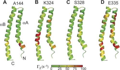
FIGURE 5.
Two time point PREs mapped on the surface of PrgI for the SipD spin labels on positions Ala-144 (A), Lys-324 (B), Ser-328 (C), and Glu-335 (D). The PREs are colored according to the strength of the Γ2 values (green being the lowest to red being the highest). The lowest energy structure of the NMR structure of PrgI that was refined by residual dipolar coupling was used.

