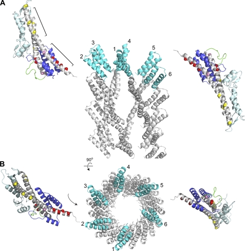
FIGURE 7.
Possible orientation of SipD in the assembled needle-tip complex as constrained by PRE results and the structure of the PrgI needle (32). Because the lower half of the SipD coiled-coil (marked by red spheres) is the primary binding site for PrgI, SipD would have be oriented in the assembled needle-tip complex with the mixed α/β domain pointed away from the needle. The reverse orientation, where the α/β domain is pointed toward the needle, will result in steric clash. The cryoEM-based model of the PrgI needle was made using coordinates obtained from Drs. Vitold E. Galkin and Edward H. Egelman (32); A and B are orthogonal views. PrgI molecules (numbered 1–6) in the topmost layer of the needle are colored cyan.

