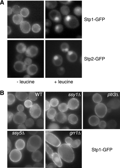
FIGURE 6.
Differential localization of Stp1 and Stp2. A, fluorescence microscopic analysis of intracellular localization of Stp1-GFP and Stp2-GFP. stp1Δ mutant cells (RBY881) expressing STP1-GFP (pZL2813) and stp2Δ mutant cells (RBY903) expressing STP2-GFP (pZL2849) were grown in SD medium ± leucine and GFP fluorescence images were captured as described under “Experimental Procedures.” B, plasma membrane localization of Stp1-GFP does not require Ssy1, Ptr3, Ssy5, or Grr1. Wild-type (ZLY044) and isogenic ssy1Δ (ZLY1915), ptr3Δ (ZLY1917), ssy5Δ (ZLY1939), and grr1Δ (ZLY1942) mutant strains expressing STP1-GFP were grown in SD medium and GFP fluorescence images were captured.

