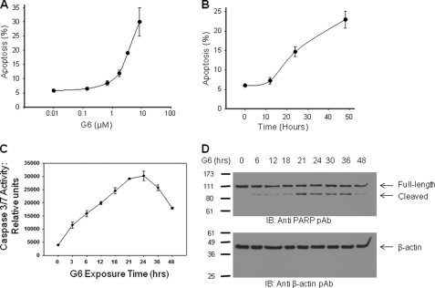
FIGURE 3.
G6 induces the intrinsic apoptotic pathway in HEL cells. For apoptotic measurements, Annexin V/propidium iodide double staining was employed. The values from three independent dose response (A) or time course (B) experiments were graphed. C, caspase 3/7 activity was measured as a function of G6 exposure time. D, after exposure to G6 for the indicated periods of time, whole cell protein lysates were Western blotted with either anti-PARP (top) or anti β-actin antibodies (bottom). Shown is one of three (A and B) or two (C and D) representative experiments. For A–C, the data are presented as the mean ± S.E.

