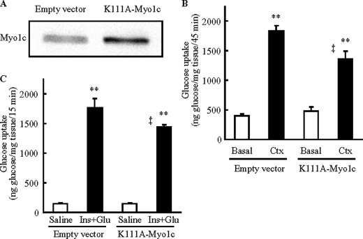
FIGURE 4.
Expression of Myo1c mutated at ATPase catalytic site (K111A-Myo1c) inhibits glucose uptake in vivo in skeletal muscle. Mouse tibialis anterior muscles were transfected with DNA vectors containing either Myo1c mutated at Lys111 to Ala (K111A-Myo1c), which has been shown to inactivate the ATPase activity of Myo1c, or the empty vector as control (Empty vector). One week after transfection, muscles were analyzed. A, muscles were harvested to assess Myo1c protein expression. B and C, mice were anesthetized, and then muscles were stimulated to contract in situ for 15 min (Ctx, B) or stimulated by injection of insulin (16.6 units/kg of insulin) and a glucose bolus to elicit maximal insulin effect (Ins+Glu, C), and tracer (2-[3H]deoxyglucose) was injected via the orbital vein. Muscles were harvested to assess 2-[3H]deoxyglucose uptake 45 min (B) or 15 min (C) after tracer injection. Open bar = basal muscle; black bar = stimulated to contract or stimulated by insulin + glucose injection. Values are means ± S.E., **, p < 0.01 versus Basal or Saline, ‡, p < 0.01 versus empty vector. The images are representative of 8 muscles (A). n = 19–20 (empty vector), 11–12 muscles/group (K111A-Myo1c) (B), 6 muscles/group (Saline), and 12 muscles/group (Ins+Glu) (C).

