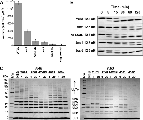
FIGURE 2.
DUB activities of the human Josephin domains: ataxin-3 (Atx3), ATXN3L, Josephin-1 (Jos1), and Josephin-2 (Jos2). A, shown is cleavage of the fluorogenic substrate Ub-AMC by the four different Josephin domains under identical conditions, as described under “Experimental Procedures.” AU, arbitrary units. B, shown is the time course of Ub-His6 cleavage catalyzed by Josephin proteins. The substrate concentration is 125 μm for all experiments; the enzyme concentration used is indicated on the left. C, cleavage of unanchored Lys-48- and Lys-63-linked ubiquitin chains is shown. Molecular weight markers are shown in the left-most lane of each gel, with corresponding molecular weights being indicated at the left. The positions of ubiquitin monomers (Ub1), dimers (Ub2), and larger oligomers are indicated. The negative control (NEG) contains no DUB enzyme. Time points were taken after 4 and 20 h of cleavage. Asterisks mark the bands corresponding to the DUB enzymes.

