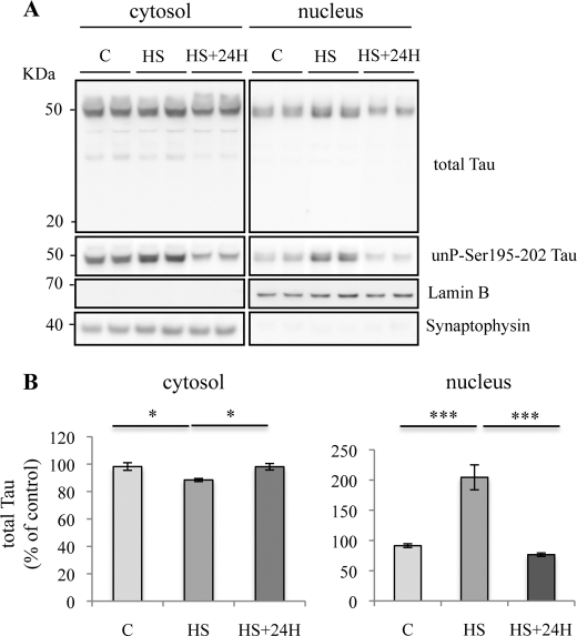
FIGURE 3.
Transient nuclear accumulation of Tau in heat stress-treated neurons. A, Western blotting analysis using phospho-independent (anti-C-terminal Tau) and dephospho-Tau (Tau1) antibodies. Cortical cultures were heated to 44 °C (HS) or kept at 37 °C (C) for 1 h and then maintained at 37 °C for 24 h. Total Tau and dephosphorylated Tau labeling were analyzed both in cytosolic and nuclear fractions. A loss of nuclear Tau staining was observed 24 h after HS. B, densitometric analysis of Western blot using anti-total Tau antibody. Results are expressed as percentage of the control. Data shown are the mean ± S.D. of three different experiments. ***, p = 0.0001; *, p < 0.05.

