FIGURE 5.
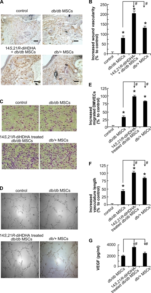
14S,21R-diHDHA remedied impaired functions of db/db MSCs and acted synergistically with them in promoting angiogenesis under hyperglycemia. The wound healing model and treatment were conducted in db/db mice as in Fig. 4. A, representative microphotographs show the CD31+ vessels in wounds treated with vehicle (control), db/db MSCs, db/db MSCs plus 14S,21R-diHDHA, or db/+ MSCs (14S,21R-diHDHA, 50 ng/wound; MSCs, 106 cells/wound). B, increased percentages of wound vascularity compared with the control that possessed 4.4 ± 0.4% vascularity (n = 12). Scale bar, 100 μm. Cellular processes of angiogenesis were studied in vitro as in Fig. 3. Representative images of db/db DMVEC migration (C) and vasculature formation (D) are shown. The db/db DMVECs were studied under simulated hyperglycemia (25 mm glucose) in DMEM (control), DMEM conditioned by db/db MSCs with (14S,21R-diHDHA-treated db/db MSCs) or without (db/db MSCs) 14S,21R-diHDHA treatment, as well as DMEM conditioned by db/+ MSCs (db/+ MSCs). Migration (E) and vasculature formation (F) were quantified as increased percentages of migrated DMVECs per field (n = 15) and of vasculature length per field (n = 4), respectively, compared with the controls (migrated DMVECs, 118.0 ± 2.8 cells/field; vasculature length, 7.2 ± 0.3 mm/field). G, VEGF secretion was assayed by Bio-Plex protein array (n = 3). The medium was conditioned with 3 × 10⁵ db/db MSCs, 14S,21R-diHDHA-treated db/db MSCs, or db/+ MSCs. Results are means ± S.E. *, p < 0.05 compared with control; #, p < 0.05 compared with db/db MSC treatment.

