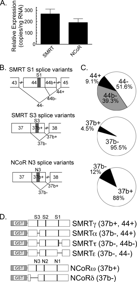
FIGURE 1.
SMRT and NCoR splice variants expressed in APL cells. A, comparable expression of SMRT versus NCoR mRNAs. Quantitative RT-PCR and splice-independent primer pairs were used to measure total SMRT and total NCoR mRNA levels in NB4 cells. The mean and S.E. of three experiments are shown. B, alternative splicing events within the SMRT and NCoR receptor interaction domains. Exons (numbered from the 5′ start site), alternative splicing events (V-shaped lines), and CoRNR motifs (S1, S3, or N3) are indicated. C, relative expression of the different SMRT and NCoR mRNA splice variants in NB4 cells. Messenger RNA isolated from NB4 cells was subjected to RT-PCR using primers spanning the splice sites in B; the products were resolved by gel electrophoresis to determine the percentage of each alternatively spliced mRNA produced at each splice site. The means of six experiments are presented. D, schematic of the GST-SMRT and GST-NCoR protein constructs. Exons deleted by alternative mRNA splicing are depicted as horizontal lines. The nomenclature is described in the text. SMRTα and SMRTτ were described previously (17, 18). SMRTγ, SMRTϵ, NCoRω, and NCoRδ were previously referred to as SMRTsp18, SMRTsp2, full-length NCoR, and RIP13Δ1, respectively (25, 46).

