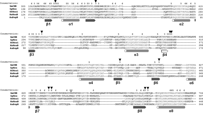
FIGURE 4.
Sequence alignment of catalytic domains of GH20 β-N-acetyl-d-hexosaminidases. Structure-based alignment was performed with PROMALS3D (31). α-Helices and β-sheets are shown in orange and blue, respectively (common ones are filled, and uncommon ones are unfilled). Important residues are indicated by triangles (common ones are filled, and uncommon ones are unfilled).

