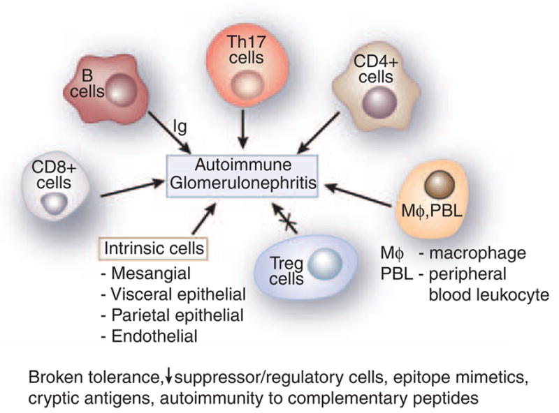
Figure 1. Cells in GN.

Figure 1 represents an aggregate of data derived from animal models. B cells were classically considered to be involved in the pathogenesis of GN by elaboration of immunoglobulin (Ig). Th-17, CD4 +, and CD8 + cells have a significant role as shown by abrogation of activity leading to amelioration of GN. Macrophages and peripheral blood leukocyte (PBL) are essential in the histological changes of GN. All result in a variable increase in the mesangial matrix, and involvement of the visceral and parietal epithelial cells. T regulatory cells downregulate disease. Experimental autoimmune glomerulonephritis presumably arises secondary to various etiologies, including broken tolerance, a decrease in suppressor/regulatory cells, epitope mimetics, exposure of cryptic antigens, and possibly autoimmunity to complementary peptides.
