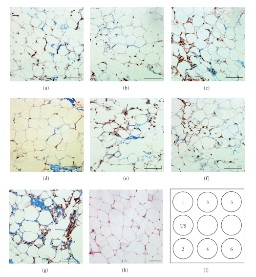
Figure 6.
Histology of the adipose organoids. The adipose organoids were cultured for 6 days with indirect LIUS stimulation for 3 minutes daily. The adipose tissue as from wells 1 (a), 2 (b), 3 (c), 4 (d), 5 (e), 6 (f), and control (g) were stained with Masson's trichrome to visualize collagen fibers (blue) and cells (red). (h) Histology of normal adipose tissue was shown as standard morphology. Schematic of LIUS setup was shown in (i). Arrows indicate stromal cells. Scale bars, 200 μm. The adipocytes from wells 1, 2, and 4 had morphology similar to normal tissue (h).

