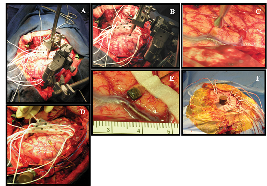
Figure 1.
Representative intraoperative photographs demonstrating elements of the microarray insertion procedure. A) Set-up of the surgical field following craniotomy, dural opening, and preliminary grid insertion. The positioning device for the impactor wand has been attached to a Greeenberg retractor system. B) Close-up view of the positioning device, demonstrating multi-dimensional capability for fine-tuning of the impactor wand. C) Close-up of the impactor wand near the cortical target, in preparation for microarray implantation. D) View of microarray following trans-pial insertion and stabilization with silk sutures to the dural edge. The device pedestal has been fixed to the skull at the edge of the craniotomy. E) Close-up of the implanted microarray, demonstrating mild sub-pial hemorrhage. F) Final appearance of the device pedestal following wound closure. In this particular patient, the pedestal was brought through the scalp flap via a separate stab incision.

