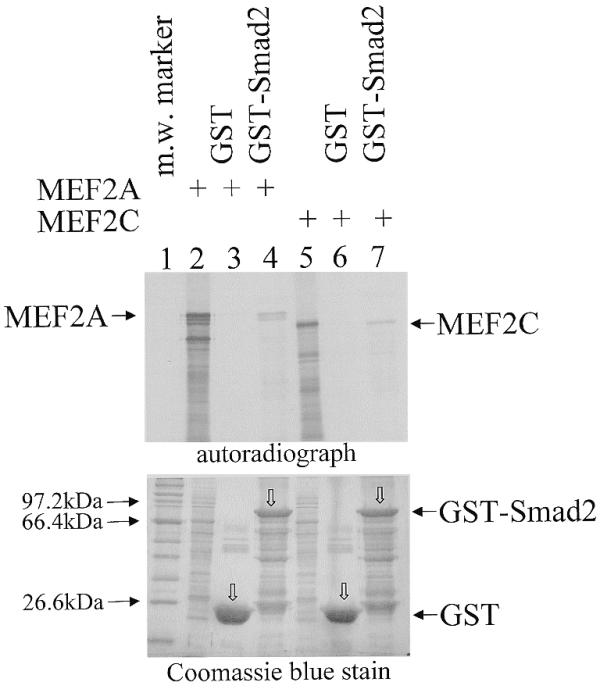
Figure 4.

MEF2 proteins associate with GST–Smad2 in vitro. (Top) In a GST pull-down assay, in vitro translated [35S]methionine-labelled MEF2A (2 µl) or MEF2C (2.5 µl) was mixed with 5 µg of GST (lanes 3 and 6) or GST–Smad2 (lanes 4 and 7) immobilised on glutathione–agarose beads. After washing, 35S-labelled bound proteins were analysed by SDS–PAGE and autoradiography. Aliquots of 0.4 µl MEF2A (lane 2) and 0.5 µl MEF2C (lane 5) in vitro translation reactions were included as references. The arrows indicate the position of MEF2A or MEF2C. (Bottom) Coomassie blue staining of the gel shows that comparable amount of the GST or GST–Smad2 proteins were loaded. The open arrows (lanes 3, 4, 6 and 7) indicate the position of the corresponding GST or GST–Smad2 fusion proteins.
