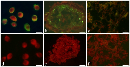
Figure 4. Localization of endogenous CfLec-1 in different tissues.
Hemocytes or 7 µm thick frozen sections of gill, adductor muscle, gonad, hepatopancreas, mantle and kidney were fixed with acetone and then incubated with rat polyclonal antiserum to CfLec-1. Binding of antibody to CfLec-1 was visualized by FITC-labeled secondary antibody (green), and the whole tissues were stained with EBD (red). a and d: hemocytes, bar = 10 µm; b and e: mantle, bar = 50 µm; c and f: gill, bar = 50 µm.

