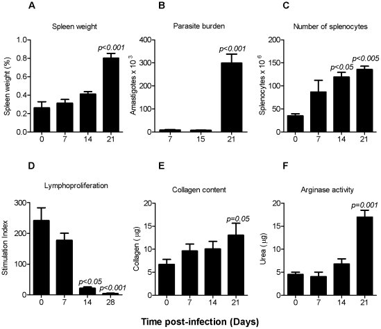
Figure 1. Rationale for selection of the time for establishing the ex vivo splenic explant culture.
Hamsters infected with 106 Luc-transfected L. donovani were evaluated from 7 to 21 days post infection (n = 6 per time point). (A) Spleen weight. Shown is the mean ± standard deviation (SD) of the spleen to body weight ratio (spleen weight divided the body weight). (B) Splenic parasite burden. The number of amastigotes (mean ± SD) was determined by luminometry in 500,000 splenocytes by extrapolating the counts (photons/sec) to a standard curve of microscopy-enumerated spleen-derived amastigotes. (C) Total splenocyte number. Splenocyte number (mean ± SD) was determined by counting the cells by microscopy. (D) Splenocyte lymphoproliferative response. The splenocyte stimulation index (shown as the mean ± SD) was determined by dividing the cpm of concanavalin A-stimulated and non-stimulated splenocytes. (E) Splenic soluble collagen content. The soluble collagen content (shown as the mean ± SD) was determined in spleens from uninfected and infected hamsters by the Sircol assay (Biocolor). (F) Splenic Arginase activity. Tissue arginase activity was determined by measurement of urea catalysis and is shown as the mean ± SD. Statistical analysis for all panels was performed by one-way analysis of variance (ANOVA).

