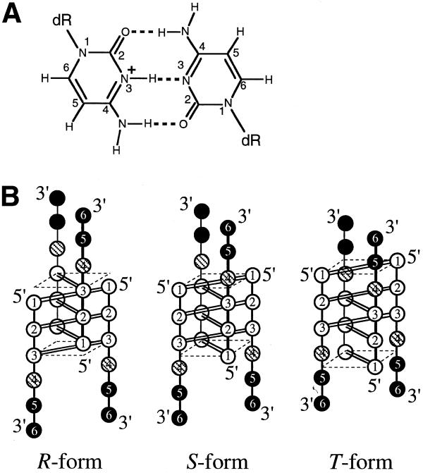
Figure 1.
(A) Chemical structure of the C·C+ base pair. dR indicates a deoxyribose moiety. (B) Schematic drawings of NMR-observable i-motif topologies of d(CCCTAA): R-form (left), S-form (middle) and T-form (right). Cytidine, thymidine and adenosine residues are symbolized by open, shaded and closed circles, respectively. C·C+ base pairs are formed in the parallel duplexes for all the topologies, indicated by double lines between open circles, and the plane formed by the outermost C1·C1+ pair is indicated by broken lines.

