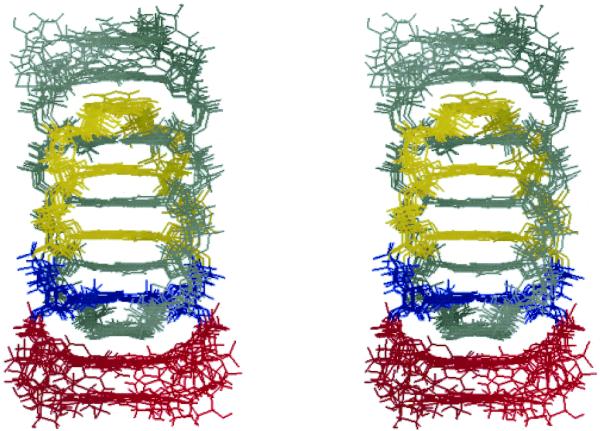
Figure 4.
Stereoview of superposition of the seven lowest-energy structures of the T-form viewed from a wide groove. C1′ atoms of C1–A5 residues were superimposed. For one parallel duplex, cytidine residues are in yellow, thymidine in blue and adenosines in red. The other parallel duplex is in gray.

