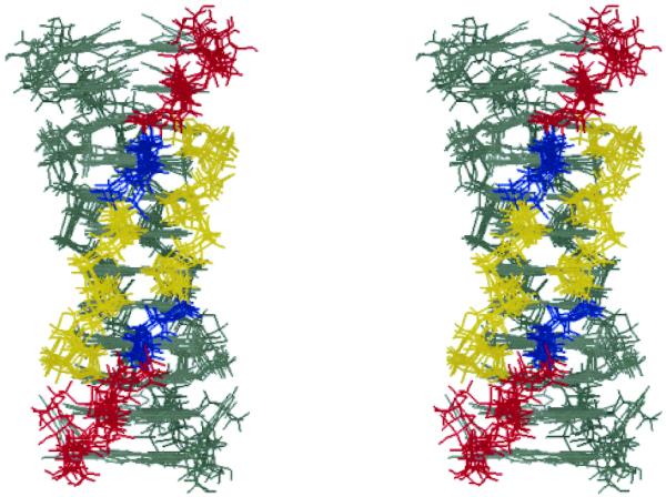
Figure 5.
Stereoview of superposition of the seven lowest-energy structures of the T-form viewed from a narrow groove. C1′ atoms of C1–A5 residues were superimposed. For one anti-parallel duplex, the bases are in gray, and the sugar–phosphate backbones are colored depending on the residue: yellow for cytidine, blue for thymidine and red for adenosine. The other anti-parallel duplex is in gray.

