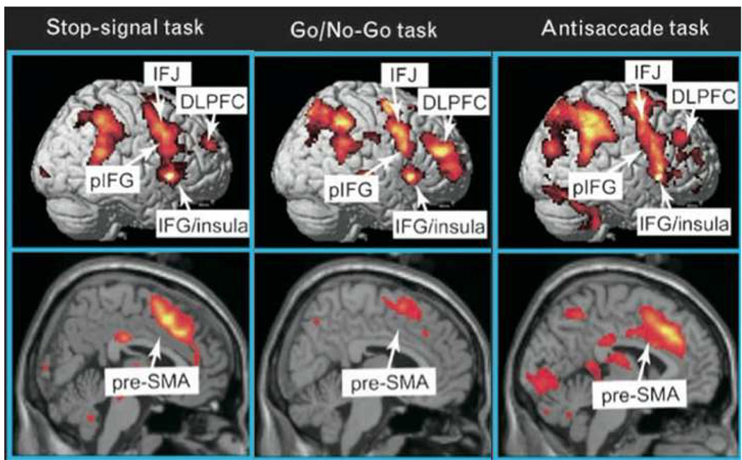
Figure 2.
Functional MRI studies of stop signal and related paradigms. The ‘stopping network’ in the cortex is activated by different control tasks and is predominantly right-lateralized. It includes the presupplementary motor area (preSMA) and the right inferior frontal cortex (rIFC). Right IFC activity is broadly distributed and may reflect an inferior frontal junction (IFJ) component, a more ventral posterior inferior frontal (pIFG) (putatively implementing inhibitory control) and an insula region of unknown function. Maps of the activation during performance of go/no-go, stop-signal and antisaccade tasks were revealed by contrasting no-go vs. frequent-go, stop vs. go, and antisaccade vs. baseline-saccade trials, respectively, see (16) for further details. Copyright permission requested.

