Figure 1.
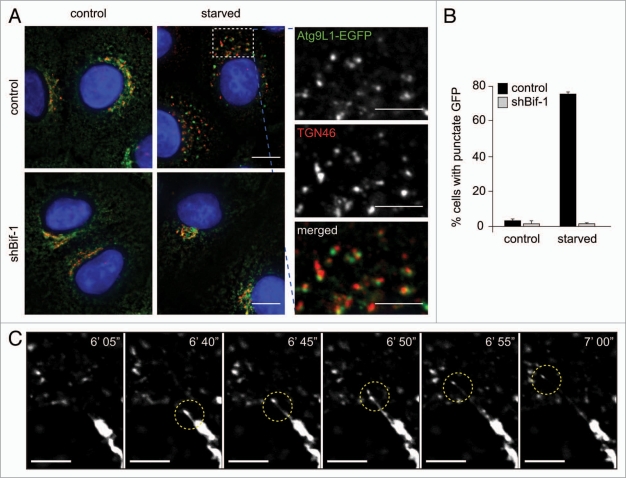
Bif-1 regulates the tubulation and fission of Atg9-positive membranes during starvation. (A) Control or Bif-1 knockdown HeLa cells stably expressing Atg9L1-EGFP were incubated in complete or starvation medium for 1.5 h. The cells were then immunostained for TGN46. The images were obtained using a fluorescence deconvolution microscope. Magnified images are shown in the right part. (B) The percentage of cells with GFP foci was calculated (mean ± SD.; n = 3 × 215). (C) HeLa cells stably expressing Atg9L1-EGFP were starved for 45 min and then analyzed by time-lapse fluorescent microscopy at 5-sec intervals. The scale bars represent 10 µm in (A), 5 µm in magnified images in (A and C).

