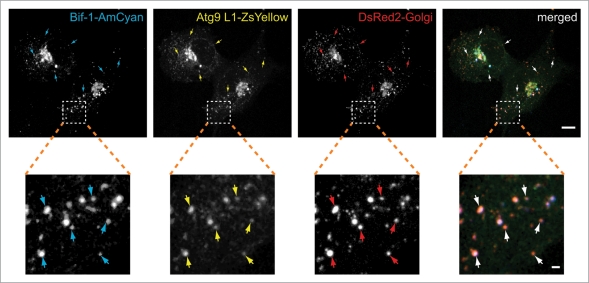
Figure 2.
Starvation-induced Bif-1 and Atg9-positive foci are derived from Golgi membranes. COS7 cells co-transfected with Bif-1-AmCyan, Atg9L1-ZsYellow and DsRed-Monomer-Golgi were cultured in starvation medium for 1.5 h. The fluorescent images were obtained using a confocal microscope. Magnified images are shown in the bottom part. Arrows indicate representative colocalization of Bif-1, Atg9 and the Golgi marker. The scale bars represent 10 µm and 1 µm in the upper and bottom parts, respectively.

