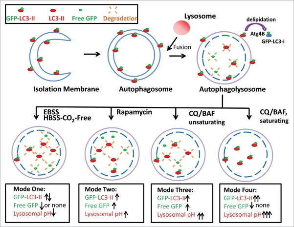
Figure 8.
Schematic diagram of the different modes of GFP-LC3 cleavage within autolysosome. Autophagy is a dynamic process. It starts from a small crescent-shaped isolation membrane, which elongates and closes to form a double-membrane autophagosome. Autophagosomes fuse with lysosomes to form mature autolysosomes, where engulfed contents including proteins and damaged organelles are degraded. During this process, LC3 is conjugated with PE at its C terminus and recruited to both the outer and inner membrane of the autophagosome. After fusion with lysosomes, the inner membrane LC3-PE is degraded by the lysosomal enzymes whereas the outer membrane LC3-PE is de-lipidated by Atg4. It is believed that GFP-LC3 may follow the same route as the endogenous LC3. However, GFP is more resistant to lysosomal degradation than LC3 and thus the free GFP fragments generated from GFP-LC3 cleavage can serve as a marker for autophagic flux. The amount of free GFP fragments can be summarized by four different modes. In mode one (rapid), few or no free GFP fragments are detected whereas the levels of endogenous LC3-II and GFP-LC3-II may fluctuate depending on the time frame of the assay. This scenario may occur during EBSS or HBSS-CO2 free-induced starvation accompanied by a decrease in lysosomal pH. In mode two (slow), levels of free GFP fragments increase as do the levels of endogenous LC3-II and GFP-LC3-II. This scenario may occur by some autophagy inducers which do not affect or only slightly increase lysosomal pH such as rapamycin or cells cultured in EBSS CO2 free conditions. In mode three (slow), the levels of free GFP fragments, as well as the levels of endogenous LC3-II and GFP-LC3-II all increase. This scenario may occur for cells or animals treated with unsaturating concentrations of lysosomal inhibitors which increase lysosomal pH either in the presence or absence of starvation. In mode four (no degradation), no free GFP fragments are detected whereas the levels of endogenous LC3-II and GFP-LC3-II are increased. This scenario may occur when cells are treated with saturating concentrations of lysosomal inhibitors which dramatically increase lysosomal pH in the presence or absence of autophagy inducers.

