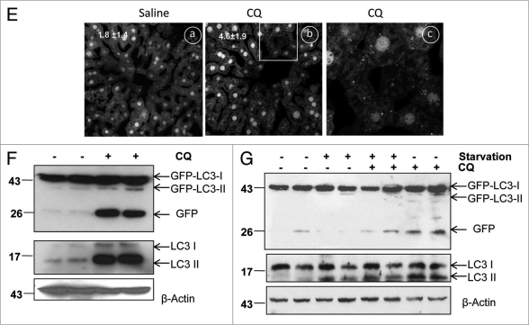
Figure 2E–G.
CQ increases free GFP fragment in various GFP-LC3 stable cell lines and GFP-LC3 transgenic mouse liver. (E) GFP-LC3 mice were starved for 6 hrs then treated with saline or CQ (60 mg/kg, i.p.) for another 16 hrs with free access to the food chow. Cryosections of GFP-LC3 transgenic livers were performed followed by confocal microscopy. Representative images are shown. a: saline, b: CQ and c: magnified image from the boxed area in b. The number of GFP-LC3 dots per cell in each condition was quantified. Data (mean ± SE ) are representative of 3 control mice and 4 CQ-treated mice from a total of more than 110 different cells. (F) After treatment with CQ as in (E), total liver lysates were prepared and subjected to immunoblot analysis. (G) GFP-LC3 mice were either starved or fed for 24 hrs with or without an injection of CQ (60 mg/kg) at the last 6 hrs before they were sacrificed. Total liver lysates were prepared and subjected to immunoblot analysis.

