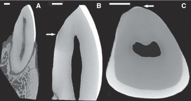
Fig. 2.

Micro-CT of the central incisors: (A) Axial view of the incisor in the bone; (B) Close-up of the axial view of the extracted incisor; note the thin and scant enamel on the lingual aspect of the tooth as indicated by the arrow; (C) Transverse view of the extracted incisor. Scale bar: 1 mm.
