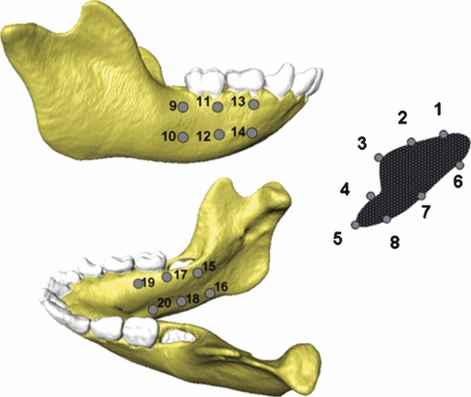
Fig. 3.

Locations of strain recordings on the FE models. ε1, ε3 and γmax strain magnitudes and the orientation of ε1 and ε3 for the PDL and the No PDL models were computed from all 20 strain locations. ε1, ε3 and γmax strain magnitudes and the orientation of ε1 and ε3 for the Crypt and No Crypt models were only computed from the symphyseal strain locations (1–8).
