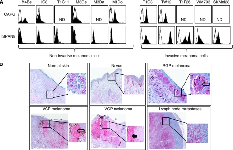
Figure 3.
TSPAN8 is expressed by invasive melanoma cells in culture and in melanoma lesions. (A) Non-invasive (M4Be, M3Ge, M3Da, M1Do cell lines and IC8, T1C11 clones) and invasive melanoma cells (WM793, SKMel28 cell lines and T1C3, TW12, T1P26 clones) were cell surface stained with antibodies directed against TSPAN8 and CAPG. Filled histograms represent specific and open histograms isotype-matched control antibodies. Results are representative of three independent experiments. (B) Representative immunohistochemical expression of TSPAN8 immunostaining in normal skin, benign nevus, RGP and VGP melanomas, and lymph node metastases. Note negative staining of cells from normal skin, except for eccrine glands, which was useful as an internal positive control. The square represents the area of magnification shown in the inset. Open arrows pointing at positive stained junctional nests of melanocytes. Black arrow pointing at a dermal nest of stained cells.

