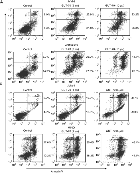
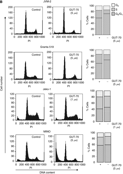
Figure 2.
GUT-70-induced cell growth inhibition, apoptosis, and cell cycle arrest in MCL. (A) JVM-2, Granta 519, Jeko-1, and MINO cells were treated with the indicated concentrations of GUT-70 for 48 h, and the percentages of apoptotic cells were quantified by annexin V/PI staining. (B) Representative flow cytometric histograms of PI-treated cells after 24 h of GUT-70 treatment at indicated concentrations. The percentages of G0/G1-, S- and G2/M-phase cells were assessed in total viable cells (bar graphs).


