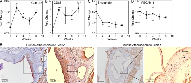
Figure 1.
GDF-15 is progressively expressed in atherosclerotic lesions in a pattern similar to that of macrophages. (A–D) Temporal expression of GDF-15 (A), CD68 (B), Smoothelin (C) and PECAM-1 (D) during atherogenesis was assessed by whole genome microarray. Values are expressed as fold induction compared with time point zero. The experiment was performed twice, with n = 3 (each containing pooled plaque material of three mice) per time point. *, P < 0.05; ***, P < 0.001, compared with the 0-wk timepoint. Error bars are depicted as SEM. (E and F) Immunohistochemistry for GDF-15 in human (E) and murine (F) atherosclerotic lesions. Arrows represent intimal cells (based on nuclear staining) that express GDF-15.

