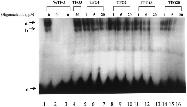
Figure 4.
Electrophoretic mobility shift analysis of the effect of TFOs on the binding of nuclear factors present in PMA-treated K562 cells to the target region of the c-sis/PDGF-B promoter. Radiolabeled duplex target, the 255 bp promoter fragment isolated from the pUC18promoter, (30 nM) was preincubated with increasing concentration (1, 5 or 20 µM) of single-stranded TFO1 (lanes 5–7), TFO2 (lanes 8–10), TFO18 (lanes 11–13), TFO20 (lanes 14–16 ), 20 µM TFO3 (lane 4) and no TFOs (lanes 1-3). Samples were then incubated with the nuclear extracts from PMA-treated K562 cells except in lane 2, which contained labeled duplex only. In lane 3, 20-fold more unlabeled duplex target was added. a and b, protein–DNA complexes; c, free duplex probe.

