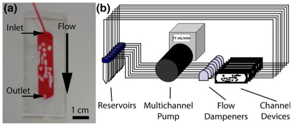
Fig. 2.
Experimental setup. (a) Microfluidic chamber replicating the channels found within a modular tissue-engineered construct. Red fluid fills the channels where endothelial cells were to be seeded. The image was taken prior to epoxy application and only shows the inlet tube. Scale bar=1 cm. (b) Eight flow circuits were set up in parallel using a multichannel peristaltic pump. Twelve mL of endothelial cell medium was circulated from separate reservoirs and was changed every 2 days. Flow dampeners were used to reduce the pulsation, eliminate backflow and also served to remove air bubbles. The entire system was placed in an incubator set at 37°C and 5% CO2

