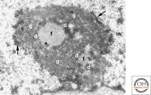
Figure 3.
The tripartite organization of the nucleolus. Electron micrograph of a mouse fibroblast nucleolus. (f) fibrillar center, (d) dense fibrillar component, (g) granular component, (arrows) perinucleolar heterochromatin, (*) denotes the presence of dense fibrillar component material within the fibrillar center, which is occasionally observed. (Reprinted from Trends in Cell Biol 13: 517–525, Raška, I. © 2003, with permission from Elsevier.)

