Figure 1.
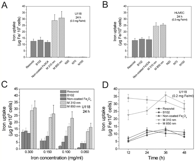
Internalization of different iron oxide nanoparticles (see Table 1) in human U118 glioma cells (A, C, D) and human umbilical vein endothelial cells (HUVEC, B) after 24 h at an iron concentration of 0.3 mg/mL; (A, B) The microspheres (0.31 μm and 0.85 μm) were absorbed best, Resovist, B102 and non-coated Fe3O4 showed an equally strong high uptake, whereas NanomagN20, N70 and N100 were not measurably internalized; (C) Iron uptake after 24 h as a function of the iron/particle concentrations applied; (D) Different uptake kinetics were observed for the different particles at the same iron concentration (0.2 mg Fe/mL); however, the strongest uptake was mostly observed between 24 and 36 h; (A–D) n = 3 ± S.D.

