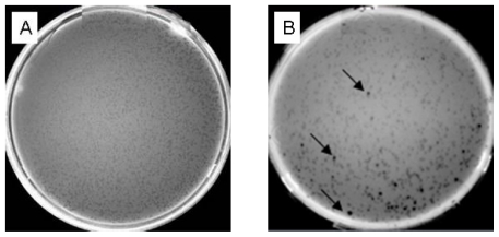
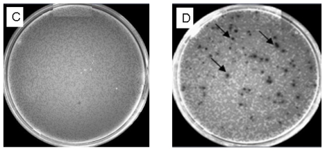
Figure 5.
(A) The titer of unamplified library. (B) Blue and white screening of the unamplified library. Arrows indicate blue plaques. The two plates (A, B) were inoculated with a 10-fold dilution of packaged phage. Plaques were approximately 1 mm in diameter. (C) The titer of amplified library. (D) Blue and white screening of the amplified library. Arrows indicate blue plaques. The two plates (C, D) were inoculated with a 1 × 10⁴ dilution of phage lysate. Bacteriophage: λ lcI857 Sam7. Host: Escherichia coli LE392MP.


