
Figure 2.
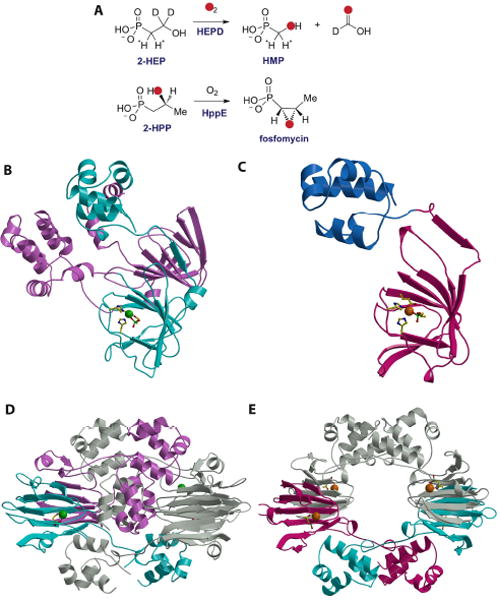
(A) Reactions catalyzed by HEPD and HppE. (B) Structure of the HEPD-HEP complex with the active metal-containing domain colored in cyan and the vestigial domain colored in pink. The bound Cd(II) is shown as a green sphere, with protein ligands and the substrate molecule shown as stick figures. (C) Structure of the HppE-fosfomycin complex with the cupin domain colored in cyan and the novel alpha domain colored in blue. The requisite Fe(II) is colored in orange with protein ligands and the substrate molecule shown as stick figures. (D) Structure of the HEPD dimer suggesting the relevance of the vestigial repeat in forming the composite active site. (E) Structure of the HppE tetramer shown in the same orientation as that for the HEPD dimer.