
Figure 4.
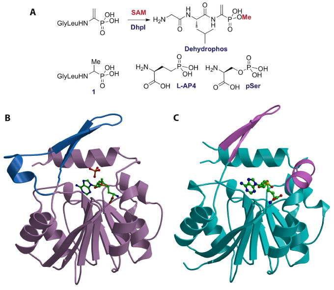
(A) Reaction catalyzed by DhpI and structures of several of the phosphonates that are also substrates for the enzyme. (B) Structure of the DhpI-SAM-sulfate complex showing the nucleotide binding Rossman fold in brown and the unusual insertions necessary for substrate binding in blue. The SAM co-factor and sulfate anion are shown as stick figures. (C) Structure of the DhpI-SAH complex with the nucleotide-binding domain colored in cyan and part of the novel insertion as well as a newly formed helix colored in pink. The helix of the insertion is disordered in the SAH complex.