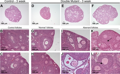
Fig. 3.
Ovary histology in 3-wk C1galt1/Mgat1 double-mutant females. Representative ovary sections from 3-wk control (A–C) and double-mutant (D–L) females contain follicles at all stages of development. Numerous follicles exist in control (A) and ovaries with double-mutant oocytes (D–F), but the size of the double-mutant ovary varied more than controls. The sections shown had the largest diameter observed in each ovary. Normal follicles in control (B and C) and ovaries with double-mutant oocytes (G and H) are shown. Immature ovaries with double-mutant oocytes contained many aberrant follicles including MOFs (I), large antral follicles with a thin granulosa layer and zona-free oocytes (J), mutant follicles fusing (K) as indicated by the visible basal lamina and theca cells between the oocytes (arrow), and follicles with granulosa cells beneath the zona pellucida (arrow) (L).

