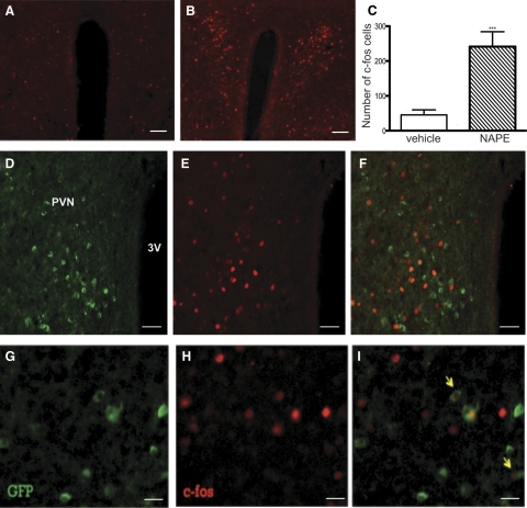
Fig. 5.
Activation of MC4R-GFP neurons by exogenous C16:0 NAPE administration. IHC of c-fos (red) in the PVN of overnight fasted male MC4R-GFP mice followed by either (A) vehicle or (B) 500 mg/kg C16:0 NAPE ip injection. Scale bar, 100 μm. C, NAPE increased c-fos–positive cells (***, P < 0.001, t test). GFP (green; D) and c-fos (red; E) positive cells and (F) double immunostaining of GFP and c-fos in the PVN are displayed. Scale bar, 50 μm. Panels G–I show higher-magnification images of double-fluorescent IHC for MC4R-GFP and c-fos. Scale bar, 10 μm. Arrows point to neurons showing double immunostaining.

