
Figure 4.
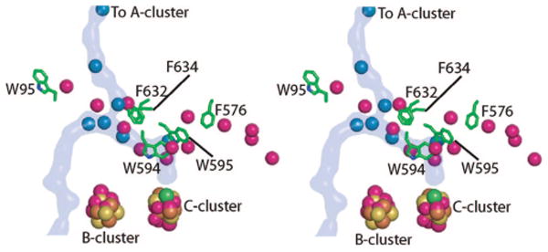
Stereoview comparing Xe sites in M. thermoacetica CODH and HCP. Xenons in the CODH structure are shown as blue spheres, and the CODH/ACS channel is highlighted in blue. The HCP xenons are colored magenta. Aromatic residues in CODH that block channels observed in HCP are shown as green sticks. Metal clusters from HCP (atoms of cluster shown as magenta spheres) align with those from mtCODH (colored by atom with iron in brown, sulfur in yellow, and nickel in green).