
Figure 5.
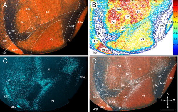
Regional pattern of neurofilament protein (SMI-32) expression in layer 4 of adult mouse cerebral cortex. A, Dark-field image of SMI-32-immunolabeled tangential section showing the posterior half of the left cerebral cortex. The gold-colored labeling shows strong SMI-32 expression in V1 (note that the sections are cut transversely at the posterior pole, which exposes the weakly labeled upper layers), Au, and S1, as well as in RSA and MEC. Moderate SMI-32 expression is found in the cortex between S1 and Au, which contains S2, DP, and DA. Moderate labeling is also observed in LEC and throughout the belt on the lateral side of V1. Much weaker expression is seen in the acallosal region on the medial side of V1. Weak labeling is also found at the lateral (ventral) tip of the belt in a region that corresponds to area 36p. Extremely sparse SMI-32 expression is present in an L-shaped region in TE and the perirhinal areas 36 and 35 on the lateral side of Au. Little detectable SMI-32 expression is seen in a longitudinal MM strip adjacent to RSA. B, Density contour map of SMI-32 expression providing a quantitative image of the staining shown in A. C, Fluorescence image of retrogradely bisbenzimide-labeled callosal connections in the same section shown in A. D, Overlay of SMI-32 labeling shown in A with white, false-colored callosal connections shown in C. The SMI-32-expressing belt around V1 is shown in an overlay of the fixed pattern of callosal connections. Callosal landmarks were used as reference for identification of areas V1, P, POR, LM, LI, AL, RL, A, AM, and PM, which were previously described by topographic mapping (Wang and Burkhalter, 2007). Labeling in P is nonuniform because of transverse sectioning of weakly labeled upper layers. Labeling of the belt's most lateral tip is weaker and outlines the weakly topographic area 36p (Wang and Burkhalter, 2007). In the rest of the uniformly callosally connected cortex, SMI-32 expression is found in a region that extends from the posterior/dorsal corner of Au into the gap between S1 and Au. This region includes DP, DA, and S2. Very sparse SMI-32 staining is shown in the L-shaped belt on the posterior and lateral side of Au, which includes TE, field 36, and field 35. Extremely sparse staining is present in MM. rf, Rhinal fissure; A, anterior; M, medial; P, posterior; L, lateral. Scale bar, 1 mm.