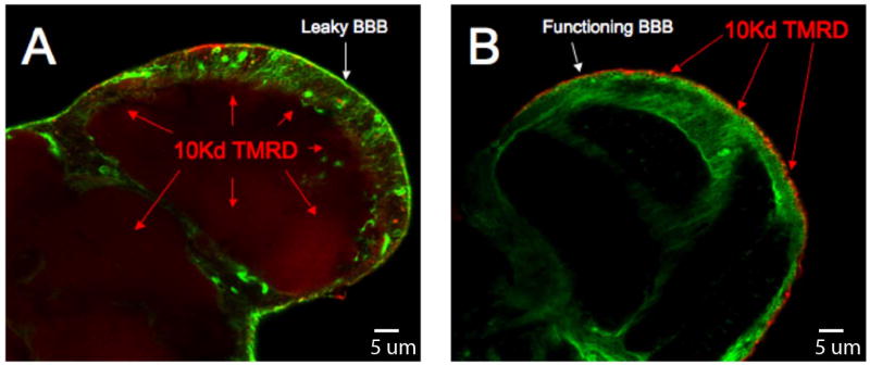
Figure E.
Confocal microscopy of the physiologic barrier to drug transport. A cross sectional confocal image of a Dm brain at the lobular plate hemolymph injected with 10 kDa tetramethyl-rhodamine dextran (TMRD). Both brain are marked with a pan glial driver (REPO-GAL4) crossed to a transgene expressing GFP (green channel) using the UAS/GAL4 system of Brand and Perrimon (23). On left the moody mutant shows strong dextran signal (red) penetrating the brain. On the right is the moody mutant rescued with Moody-GFP showing wild type BBB function. Here the dextran is fixed to surface tissue outside the brain (red).

