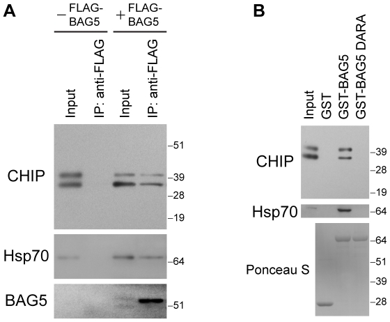
Figure 5. CHIP forms a protein complex with BAG5.
(A) Immunoprecipitations with anti-FLAG antibodies were performed from lysates of H4 cells transfected with CHIP with or without FLAG-BAG5 as indicated. Immunoprecipitates were sequentially probed with anti-CHIP (upper), anti-Hsp70 (middle), and anti-FLAG (lower) antibodies. Ten percent of lysates used for immunoprecipitation was loaded as input. The upper CHIP band corresponds to monoubiquitinylated CHIP [62]. Molecular weight markers are shown on right. Similar results were found in three separate experiments. (B) PDAs were performed using lysates of H4 cells transfected with CHIP. Proteins that associated with GST alone, GST-BAG5, or GST-BAG5 DARA were probed with anti-CHIP (upper) and anti-Hsp70 (middle) antibodies. Input was 10% of lysates used for PDAs. The presence of equal amounts of GST fusion proteins was confirmed by Ponceau S staining of the membranes (lower). Molecular weight markers are indicated on right. Results are representative of four independent experiments.

