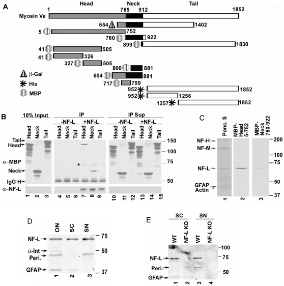
Figure 1. Schematic representation of Myo Va deletion constructs.
(A). Amino acid numbers correspond to the mouse dilute gene product [6]. The three domains of Myo Va are color coded (head-grey; rod-black and tail-white). The mutant proteins are either tagged with MBP (Hatched-circle) or hexa-His-peptide (star) or 3-kDa β-gal peptide (hatched-triangle). Constructs 5–752, 760–922 and 899–1830 are from chicken [11]. The construct tagged with β-gal-654-1402 is of human origin [15], and the tail clones originate from the mouse sequence [6]. Clones 718-799 and 800–881 are PCR products from chicken Myo Va [11]. (B) Relative binding of Myo Va tail, head and neck regions to NF-L. Myo Va head, neck and tail domains were incubated with NF-L, co-immunoprecipitated with NF-L antibody (NR-4), immunoprecipitates (IPs) and supernatants (Sup, 10%) were immunoblotted with anti-MBP and NF-L antibody (NR-4) and immunoreactive bands were detected with ECL reagent. The molecular weight (MW) of Myo Va head domain-120, neck-60 and tail is 140-kDa. *-indicates non-specific binding. (C) Myo Va head region shows more binding to NF-L compared to the neck region in blot overlay assays. Cytoskeletal fractions from spinal cord fractionated on 7% acrylamide gels containing SDS, transferred to nitrocellulose membranes, blocked with 3% milk, incubated with head and neck constructs tagged with MBP at equimolar concentrations, immunoblotted with anti-MBP-HRP antibody and immunoreactive bands were detected with ECL reagent. Ponc. S: Pnoceau S. (D) Myo Va head region binds to NF-L, α-internexin, peripherin and GFAP in blot overlay assays. Cytoskeletal fractions from optic nerve (ON), spinal cord (SC), and sciatic nerve (SN) were fractionated, and the Myo Va head domain binding activity was detected by blot overlay assay as described in panel C. (E) Myo Va head region binding to NF-L is specific since NF-L deficient cytoskeletal preparations do not show NF-L binding activity. Cytoskeletal fractions from WT and NF-L null (NF-L KO) mice spinal cord (SC) and sciatic nerve (SN) were fractionated and blot overlay assays were performed to detect Myo Va head domain binding activity as described in panel E. The positions of the bands on the membranes are indicated with arrows in all panels. Anti-MBP: α-MBP; anti-NF-L:α-NF-L.

