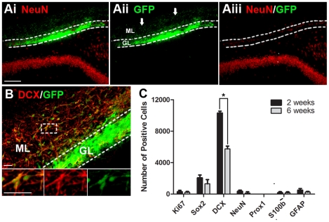
Figure 3. Transplanted primary vSVZ cells fail to become DG granular neurons in-vivo.
vSVZ cells were isolated from neonatal GFP rats and transplanted immediately after isolation. (Ai–Aiii) Example of a GFP-positive vSVZ cell transplant in the hippocampus 6 weeks after grafting. Note that the transplanted cells did not repair the damage in the granular layer (GL). Note also that many of these DCX+ cells have migrated beyond the granular layer into the molecular layer (ML). (B) Example of grafted cell expressing the immature neuronal marker DCX. (C) Total number grafted cells per animal expressing different markers at 2 and 6 weeks (mean ± SEM, n = 4), *p<0.05. Note that few cells expressed NeuN and no cells expressed Prox1. Scale bar = 100 µm for A; scale bar = 50 µm for B.

