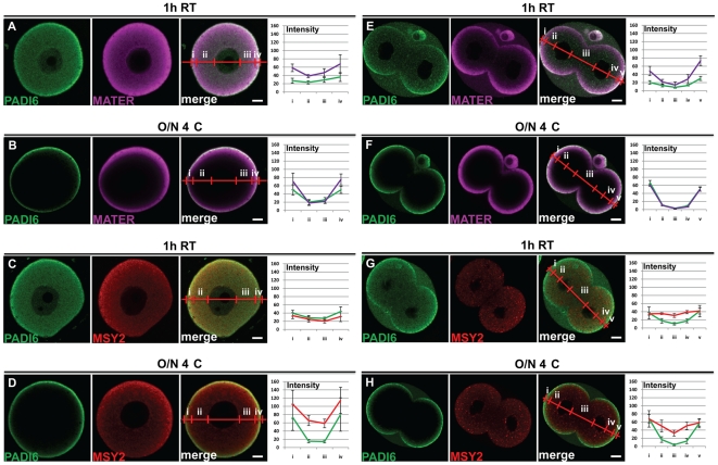
Figure 1. Time and temperature influence the staining pattern of PADI6 and MATER, but not MSY2.
GV oocytes (A-D) and two-cell embryos (E-H) were prepared for IF and stained with antibodies against PADI6 (A-H), MATER (A, B, E and F) or MSY2 (C, D, G and H). Primary antibody incubation was carried out at either 1 h RT (A, C, E and G) or O/N 4°C (B, D, F and H). PADI6 is shown in green, MATER in magenta and MSY2 in red. Included are graphs of the averages and standard deviations of four intensity profiles of four images per condition of the different stains in either cortical (i and iv) or cytosolic (ii and iii) zones for the oocytes or cortical (i and v), basolateral cytosolic (iii) or apical cytosolic (ii and iv) zones for the embryos. Bars, 10 µm.

