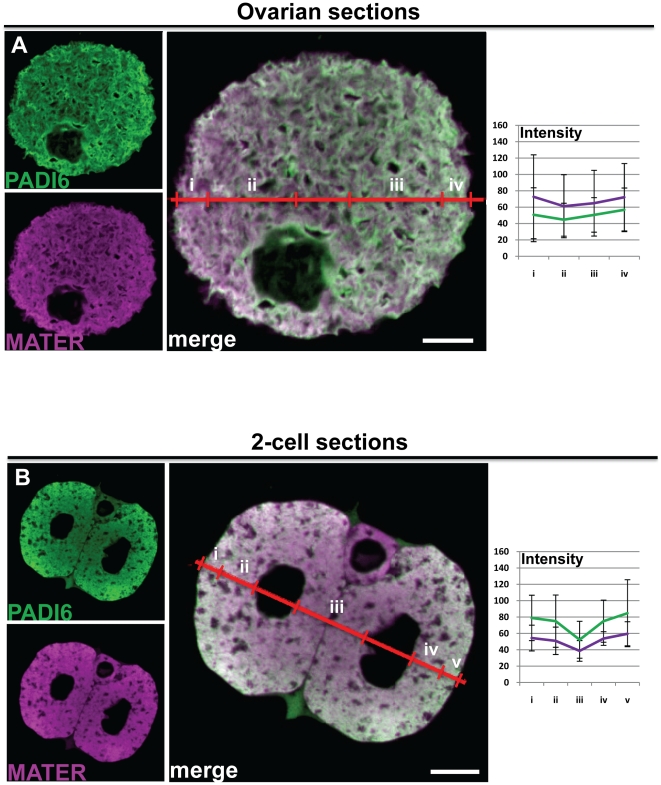
Figure 4. IF of cross sections reveal that PADI6 and MATER are primarily localized to the cytoplasm.
Ovaries and oviducts were fixed, embedded in paraffin and sectioned before staining for IF. GV oocytes (A) and two-cell embryos (B) in the sections were visualized. PADI6 is shown in green and MATER in magenta. Included are graphs of the averages and standard deviations of four intensity profiles of four images per condition of the different stains in either cortical (i and iv) or cytosolic (ii and iii) zones for the oocytes or cortical (i and v), basolateral cytosolic (iii) or apical cytosolic (ii and iv) zones for the embryos. Bars, 10 µm.

