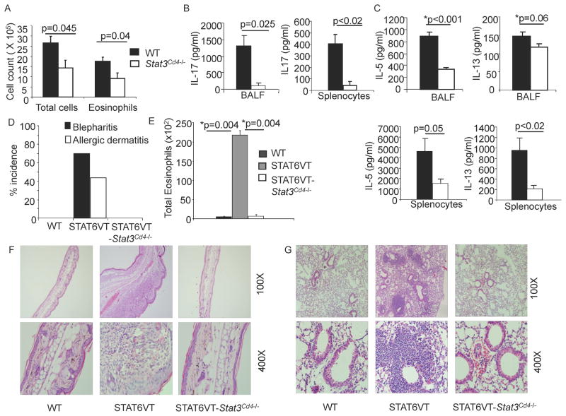
Figure 6. STAT3 Promotes the Development of Allergic Inflammation.
(A–C) WT and Stat3Cd4−/ − mice were immunized with OVA and Alum on days 0 and 7 and challenged as described in methods. After challenges, BAL cell numbers were determined by cell counting and flow cytometry (A), and cytokine levels were measured in BAL fluid and in culture supernatants from splenocytes stimulated with OVA for 72 h using ELISA. Data are average of 5–6 mice per group ± s.e.m
(D) Incidence of blepharitis and atopic dermatitis of WT, STAT6VT and STAT6VT-Stat3Cd4−/ − mice are shown. Incidence was determined by visual examination of mice (n=25 per group).
(E) Numbers of eosinophils (defined by flow cytometry) recovered in BAL. BAL data are representative of 2 independent experiments and shown as the average of 2 mice per group ± s.d. For A–C and E, Students t test was performed to calculate p values.
(F) Ear tissue from WT, STAT6VT and STAT6VT-Stat3Cd4−/ − mice were fixed and paraffin-embedded sections were stained with hematoxylin-eosin. Magnification is indicated in the panel and photomicrographs are representative of 10 mice per group
(G) Lungs from WT, STAT6VT and STAT6VT-Stat3Cd4−/ − mice were embedded in paraffin and stained with H & E. Magnification is indicated in the panel and photomicrographs are representative of 10 mice per group.

