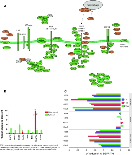
Fig. 4.
a Loss of autocrine RTK signaling networks through EGFR/ErbB2/ErbB3, Met/Ron and IGF1R signaling networks. Attenuated IL4R, Muc1 and Muc4 and integrin α6β4 components also were observed. b Attenuated RTK tyrosine phosphorylation in H292 and H358, (epithelial phenotype) Calu6 and H1703 (mesenchymal phenotype) cell models were confirmed by quantitation of RTK arrays. c EGFR is functional and responds to exogenous ligand in both epithelial and mesenchymal states. H292 and H358, (epithelial) Calu6 and H1703 (mesenchymal) cells were exposed to EGFR kinase inhibitor (3 μM erlotinib, 2 h) or DMSO control prior to stimulation with exogenous EGF (10 ng/ml, 10 min). The EGFR-dependent inhibition of substrates CBL and SHC were similar in epithelial and mesenchymal-like cells, relative for control HSPD1

