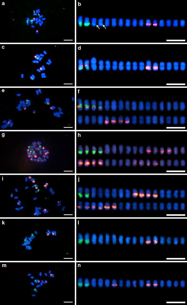
Fig. 1.

Fluorescence in situ hybridization of mitotic metaphase spreads (a, c, e, g, i, k, m) and chromosome alignments (b, d, f, h, j, l, n) of Byblis aquatica (a, b), B. filifolia diploid cytotype (c, d), B. filifolia tetraploid cytotype (e, f), B. guehoi (g, h), B. liniflora (i, j), B. rorida cytotype A (k, l), and B. rorida cytotype B (m, n). 5S rDNA (red fluorescence) and 45S rDNA (green fluorescence) probes were simultaneously detected. Chromosomes were counterstained with DAPI. Arrows minor 45S rDNA signals. Bars 5 μm
