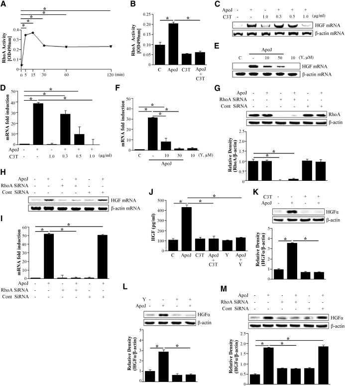
Figure 2. Activation of RhoA is required for apoptotic cell-induced HGF mRNA and protein expression.
RAW 264.7 cells were stimulated with apoptotic cells for the times indicated. (A and B) The levels of activated RhoA were determined using the RhoA activation assay Biochem Kit™ (G-LISA™). (B–F) RAW 264.7 cells were preincubated with a specific Rho inhibitor, C3 transferase (C3T; 0.3–2 μg/ml) for 20 h or a Rho kinase inhibitor Y27632 (Y; 10 and 50 μM) for 2 h before stimulation with apoptotic Jurkat cells for 2 h. (G) RhoA expression in RAW 264.7 cells transfected with RhoA siRNA or control (Cont) vehicle (siRNA-GFP) for 24 h was analyzed by Western blotting with antispecific RhoA. (H and I) RAW 264.7 cells were transfected with RhoA siRNA or control vehicle (siRNA-GFP) for 24 h and then stimulated with apoptotic Jurkat cells for 2 h. HGF mRNA levels were analyzed by semiquantitative RT-PCR (C, E, and H) and real-time PCR (D, F, and I). RAW 264.7 cells were preincubated with 1 μg/ml C3 transferase for 20 h and 50 μM Y27632 for 2 h (J–L) and transfected with RhoA siRNA or control vehicle (siRNA-GFP) for 24 h (M) and then stimulated with apoptotic Jurkat cells for 24 h. (J) HGF levels in the conditioned medium were measured by ELISA. (K–M) Western blots with anti-specific HGF α-chain were used, using the cultured cell lysates. Relative values of HGF-α expression are indicated below the gel. Values represent means ± se of three separate experiments; *P < 0.05.

